 Figure 2.1 |
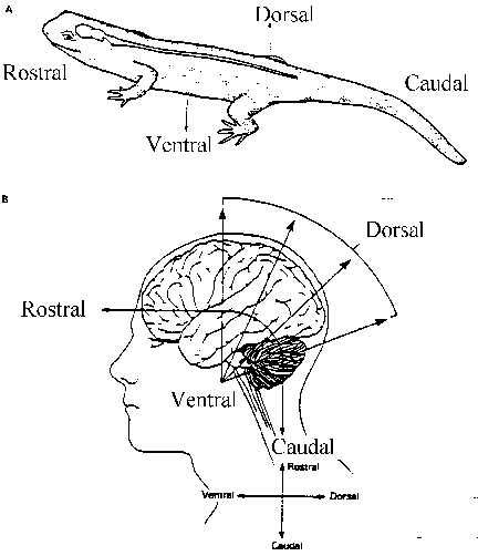
Many different terms are used to specify the relative locations of specific portions of the nervous system. Some of these terms overlap partially or even completely. Anterior and cephalic both refer to the direction towards the head end of an organism; posterior and caudal likewise both refer to the direction of the tail end of the organism. Medial is used for the direction towards the middle (on the vertical axis) of an organism, and lateral is used for the opposite direction (towards the left or right side of the organism).
 Figure 2.1 |
Caution must be exercised in the use of the pair of terms dorsal and ventral because they are used differently depending on whether one is referring to a four-legged or two-legged animal. Dorsal means "towards the surface or back"; in a four-legged animal this is always its back, but in a two-legged animal it also is used to refer to the top of the brain. Ventral means "towards the belly or front"; it refers to the bottom of a two-legged animal's brain as well (See Figure 2.1).
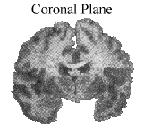
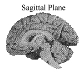
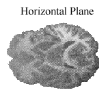
In addition to these terms which describe relative location, it is useful to know terms that specify various views of the brain. A view from the left or right side is known as sagittal; a view from the front or back is called frontal or coronal; a view from the top or bottom is called horizontal. (See Figure 2.2).
 |
 |
 |
Figure 2.2 |
||
Figure 2.3 |
In order to protect the delicate tissues of the central nervous system, there exist several layers of shielding. Most obvious to the casual viewer are the hair, which provides padding and warmth, and the skull. Inside the skull there are several meninges, or protective coverings. The outermost layer, the dura mater, is a very tough (cf. Latin durus, hard, tough) membrane. The innermost layer, the pia mater, is much softer (cf. Latin pius, tender) and adheres to and follows the contours of the brain. The arachnoid membrane, a spiderlike structure, is between the pia and dura matters. The space below the arachnoid is known as the subarachnoid space; it is filled with cerebrospinal fluid (CSF).
The clear, colorless CSF also fills a series of chambers in the brain called the cerebral ventricles. CSF is continually manufactured from blood plasma in specialized cells in the lateral ventricles called the choroid plexus. CSF leaves the brain through one-way valves called the arachnoid granulations which let CSF out into the superior sagittal sinus where it can be disposed of. Every three hours the brain’s supply of CSF is completely replaced. CSF acts mechanically as a shock absorber for the brain to protect it from sudden movements of the head. It also is the medium by which blood vessels and brain tissue exchange materials.
The delivery of nutrients and other substances to brain cells and the removal of waste products take place at very fine capillaries which branch off from small arteries. This exchange is unique to the brain in that many substances move from the capillaries into the brain cells with much more difficulty than into cells in other organs of the body. Active transport mechanisms select the substances which the brain needs. This is called the blood-brain barrier; it serves to protect the brain from exposure to some substances found in the blood. Some areas in the brain need direct access to the bloodstream and are not protected in this way. For example, the circumventricular areas, which are involved in the control of glands and hormones, are susceptible to foreign substances in the blood.
The nervous system of vertebrates has two major divisions: the CNS (comprising the brain and spinal cord) and the PNS. Notice that each of these is subdivided into various parts. In the development of an organism, these divisions become apparent at different stages.
|
Brain. The brain arises from the anterior end of the neural tube. Anatomically, the brain begins as a continuation of the spinal cord. Early on in development the neural tube changes in shape and acquires a 90 degree bend. The future brain divides into the forebrain, midbrain (mesencephalon), and hindbrain (rhombencephalon). Each of these further subdivides.
The forebrain divides into the telencephalon (which contains the cortex, basal ganglia, and limbic system) and the diencephalon (which contains the thalamus and hypothalamus). The midbrain has two major parts: the tectum (from the Latin for "roof", which contains the superior and inferior colliculi) and the tegmentum (from Latin for "floor", which contains the reticular formation). The hindbrain divides into the mesencephalon (containing the pons and cerebellum) and the myelencephalon (containing the medulla).
The fully developed mammalian brain is divided into five major lobes: frontal, parietal, occipital, temporal, and limbic. The central sulcus separates the frontal and parietal lobes, the Sylvian Fissure separates the temporal from the frontal and parietal lobes, and the parieto-occipital sulcus separates the parietal and occipital lobes. The preoccipital notch is a small depression which separates the temporal and occipital lobes.
The two hemispheres of the brain are connected by a tract of fibers called the corpus callosum. In some epileptic patients, cutting the corpus callosum can help reduce the number or severity of seizures. Researchers discovered that different portions of the corpus callosum connect different lobes of the brain, which led to advances in the sophistication of split-brain operations. The corpus callosum is divided into the genu, the body, the splenium, and the anterior commisure. The genu of the corpus callosum connects the two frontal lobes. The body interconnects the parietal lobes, while the splenium connects the occipital lobes. The anterior commissure carries fibers connecting the temporal lobes.
Spinal Cord. The spinal cord is also part of the CNS. It consists of a tiny canal surrounded by two regions of nervous tissue, the gray and white matter. Gray matter contains cell bodies and dendrites and is mainly concerned with reflex connections at different levels of the vertebral column. The white matter contains mostly axons of interneurons which are covered with myelin sheaths. Many of the axons are bundled into sensory nerve tracts which ascend the spinal cord to specific brain regions. Other axons are bundled into motor tracts that run in the opposite direction, down the spinal cord.
The other major division of the nervous system is the PNS, or peripheral nervous system. In the PNS, nerves which carry information to the brain are called afferent (from Latin ad ferre, to bear towards); nerves which carry information from the brain are called efferent (from Latin ex ferre, to bear away from). The efferent nerves are divided into two systems (see figure 2.3): the somatic and the autonomic systems. The somatic system consists of nerves leading to the skeletal system. Fibers leading to the heart, smooth muscles, and glands are the autonomic system.
The autonomic system is further divided into two categories: sympathetic and parasympathetic nerves. The parasympathetic system serves to slow down things such as heart rate and devote more energy to "housekeeping" tasks such as digestion. During times of heightened awareness, excitement, or danger, the sympathetic nerves reduce energy for housekeeping tasks and increase other activities which prepare the animal for "fight or flight."