Content created: 2006-07-14
Minor content revision: 2017-09-21
File last modified:
Human Birth & Bipedalism
An Overview for College Students
Human bipedalism is promoted by a narrow pelvis. However in mammals the pelvis is also the passage through which newborn babies pass, and the birth canal must be sufficiently large to accommodate a birth. Thus there are evolutionary pressures favoring a narrow pelvis, efficient in locomotion, conservative of bone, and less likely to break. But there are other evolutionary pressures favoring a wide pelvis, at least in women, to allow the passage of a developed fetus.
The two photos show plastic casts. In each picture, the upper two pelves are humans, female to the left, male to the right. Below them is a chimpanzee (sex unknown). Your first observation will be that chimp doesn't look at all like the humans. That's not very surprising; the chimp is not bipedal.
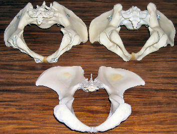
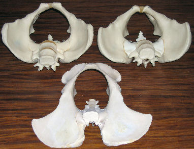
Left picture: pelves looking from outside into the body lying on its stomach. Right picture: pelves looking from inside out of the body lying on its back. In each picture the human female is top left, human male top right, and modern chimpanzee (sex unknown) at the bottom.
The picture on the left shows the pelves as seen from outside the body looking inward (with the tail bone closest to us and the body lying on its stomach). The picture on the right shows the pelves as seen from the inside of the body looking out (with the cross-section of the spinal cord facing us and the body lying on its back).
Notice how different the male and female pelves are. The male pelvis, which doesn't have to be able to pass a baby, is narrower than the female pelvis, and has a much smaller "birth canal." The casts were made from relatively "extreme" individuals in order to highlight this difference, so this is roughly the maximum difference one finds in most human populations. The genetics of sexual dimorphism are very complex, and it is not clear whether the male pelvis is "ideal" for bipedal locomotion or whether it shares some of the compromise for child passage that the female pelvis has. The female pelvis makes as little concession as possible to childbirth, but must make some.
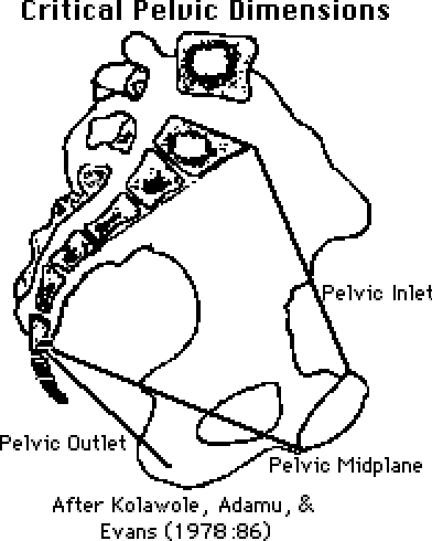
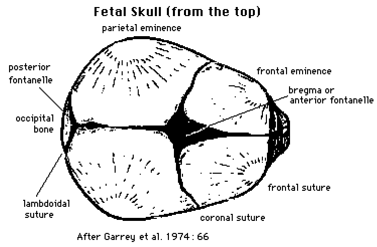
Although there are individual differences (and, naturally, classifications of the differences), for most women the narrowest place in the birth canal is the "pelvic inlet" at the top of the birth canal. The pelvic inlet is usually wider from side to side than from front to back. As the fetus descends into position for birth, it turns, as shown below, so that the head (which is longer from front to back than side to side) faces one of the mother's hips.
In most births, the baby's head is larger than the birth canal, and if there were no flexibility on either side, the baby could not be successfully delivered, and mother and baby would both usually die. Two mechanisms make human birth possible.
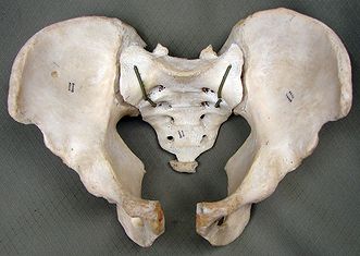
- Shortly before birth, the mother's body produces a hormone that causes the relaxation of the ligaments that hold together (1) the pubic symphysis (at the very front, bottom end of the birth canal) —a "symphysis" is the meeting point of two adjacent bones— and (2) the sacroiliac symphyses (where the bottom of the spine —the sacrum— attaches to the large, dish-shaped ilium or "hip bone" on each side of it). The technical name for this loosening is "subluxation." In extreme cases, the pubic symphysis may open a centimeter or more, making it difficult or impossible for a woman to walk in the period just before birth. However the mean is about 4 mm. Even this can produce an adequate increase in the cross-sectional area of the birth canal to allow the baby's head to pass. (For clarity, the photo here shows these symphyses more widely open than would occur in a normal birth.)
- A baby's head is somewhat flexible because the plates of bone of which it is composed
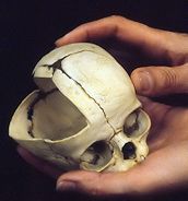 have not yet fused together at the symphyses of the skull, allowing the head to "compact" slightly with little damage in most cases. (The cut-away in the photograph shows the symphyses from both the inside and outside of the skull.) The actual flexibility of the bones themselves in the head of a fetus at full term is roughly that of adult bone, so as the bones are pushed together, they normally lock and prevent brain damage, although in some cases one plate may slide slightly below another.
As the head passes through the birth canal, the fit is nevertheless always extremely tight, with strong pressures compacting the infant's head and sometimes ripping the tissues that hold the front of the mother's pelvis together. In examining the skeleton of a woman who has delivered children, one can usually see scarring on the pubic symphysis from this process. (It is even possible to count the number of deliveries from these scars. However, it is an inexact measure. Check here for a note on false positives on another web site.)
In premature babies, although the head is smaller, the density of the fetal bone is less and the flexibility greater, producing a higher risk of brain damage from the pressure of passing through the birth canal.
As the child's head begins to emerge from the pelvic outlet, the shoulders are entering the pelvic inlet, and they too are subject to intense pressure and a narrow passage. (Consider what you would be up against if you had to get your shoulders through a space that your head could barely fit through.) At this point the fetus rotates further so that the shoulders can move through the birth canal parallel with its widest dimension. If the shoulders do not successfully emerge (a condition known as "shoulder dystocia"), the baby may be severely damaged or killed in a forced birth, or, without surgical intervention, both mother and baby may die.
Obviously, additional dangers are posed when babies emerge feet-first or when multiple fetuses get tangled with each other or prevent each other from emerging in the right position.
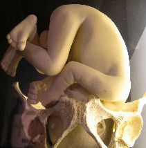
Because of the rotation of the fetus during delivery —first to turn the head, then to turn the shoulders— most children are born with their faces towards the mother's back (with the face toward the ground if the mother is lying on her back). This places the child in a very vulnerable position during the later stages of the delivery if the mother has no assistance. If the mother attempts to speed the process by pulling the fetus out with her hands, she is likely to bend its spine backwards, potentially damaging it. (Human spines bend forward quite easily. In comparison, they bend backwards almost not at all, even in neonates.) For this reason, among others, unattended births among humans are riskier than those with a midwife or other assistant present. And all births are dangerous.
(Interestingly, the English term “midwife” is etymologically derived from Middle English mid = “together with” and wyfe = “woman,” that is the woman who is beside the woman giving birth. “Obstetric” is from Latin obstāre = “to stand in front of,” that is, to stand in front of a woman as she gives birth.)
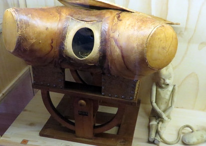
XIXth-Century Kit for Training Midwives
The cloth doll at right is the kit’s “baby,” with umbilical cord and afterbirth, to be placed inside the leather model and “delivered” by trainees through the open “vagina.”
Today mothers have the option of cesarean section, which involves cutting a hole in the abdominal wall and into the uterus and delivering the child through the hole rather than through the birth canal. (The process is named after Julius Caesar, once believed to have been delivered this way.) Before the availability of modern surgical techniques, sanitation, and antibiotics, such a process would normally have involved the death of the mother, if not immediately, then quite soon afterward. Today it is sometimes considered safer than giving birth through the birth canal.
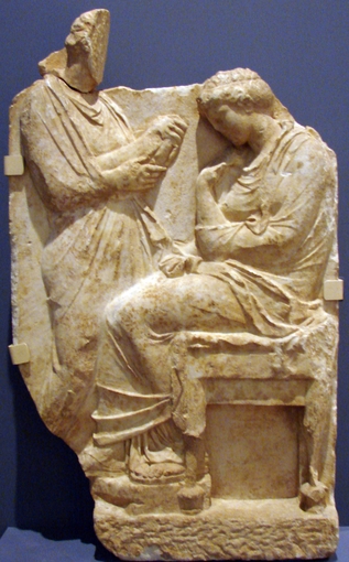
Funerary Stele for Childbirth Death
The woman at the right died in childbirth. The figure at the left is a relative holding the surviving newborn. (Athens, IVth-Century BC)
The human infant is always born quite immature, utterly helpless, and not particularly quick (compared with other animals) to attain even minimal approximations to adult competence. In the animal world in general, longer gestation periods (periods in the womb) are correlated with shorter time to maturity after birth (once one takes account of variations in body size). It is usually argued that the human gestation period is shorter than it "should" be because if the child remained longer in the womb and became a larger fetus, with an larger head, then the birth canal would have to be too large to allow functional bipedalism, at least in women.
In other words, the complexity, pain, and danger of human birth and the long and dangerous dependency of the human infant are the "price" we pay for hips that facilitate bipedal locomotion. What was there about being bipedal at the time when bipedalism first developed that offered an adaptive advantage great enough to offset these obvious disadvantages? That is a question that has attracted much speculation, but about which experts continue to disagree.
A review quiz is available for this page:
Quiz
Sources
This page is a by-product of an article on Neanderthal pelvic anatomy I wrote with my colleague Nancy Friedlander in 1995. That article is listed below, together with the sources most directly relevant to the matters discussed above.
-
AIELLO, Leslie & Christopher DEAN
- 1990 An introduction to human evolutionary anatomy. London: Academic Press.
-
FRIEDLANDER, Nancy & David K. JORDAN
- 1995 Obstetric implications of Neanderthal robusticity and bone density. Human Evolution (Florence) 9: 331-342.
-
GARREY, Matthew M., A.D.T. GOVAN, Colin HODGE, Robin CALLANDER
- 1974 Obstetrics illustrated. Second edition. Edinburgh: Churchill Livingstone.
-
KOLAWOLE, T.M., S.P. ADAMU, & K.T. EVANS
- 1978 Comparative pelvimetric measurements in Nigerian and Welsh women. Clinical Radiology 29: 85-90.
-
LEUTENEGGER, Walter
- 1982 Encephalization and obstetrics in primates with particular reference to human evolution. IN Este ARMSTRONG & Dean FALK (eds.) Primate brain evolution: methods and concepts. New York: Plenum Press. Pp. 85-95.
- 1987 Neonatal brain size and neurocranial dimensions in Pliocene hominids: implications for obstetrics. Journal of Human Evolution 16: 291-296.
-
OHLSÉN, Hans
- 1973 Moulding of the pelvis during labour. Acta Radiologica Diagnosis 14: 417-434.
-
PUTSCHAR, Walter G.J.
- 1976 The structure of the human symphysis pubis with special consideration of parturition and its sequelae. American Journal of Physical Anthropology 45: 589-594.
- The photo of the fetus placed in the pelvic girdle is from an exhibit at the San Diego Museum of Man. The cut-away fetal skull is also from the museum's collections. The midwife-training kit is from the University Museum, Utrecht, Netherlands. The funerary stele is from the Musée des Beaux-Arts, Montréal. The pelves belong to the Department of Anthropology, UCSD.
Return to top.
This page has received

visits since 160918.
 have not yet fused together at the symphyses of the skull, allowing the head to "compact" slightly with little damage in most cases. (The cut-away in the photograph shows the symphyses from both the inside and outside of the skull.) The actual flexibility of the bones themselves in the head of a fetus at full term is roughly that of adult bone, so as the bones are pushed together, they normally lock and prevent brain damage, although in some cases one plate may slide slightly below another.


